PARADIM Highlight #55—Education and Outreach (2022)
PARADIM Data Collective
The Materials Innovation Platform PARADIM maintains the PARADIM Data Collective (PDC) to make data and software that are associated with publications by Platform users publicly available. Among all available datasets, electron microscopy data had the most downloads from unique non-bot users. Further below we highlight three examples and the associated original publications.
PARADIM is establishing the utility of new imaging tools (e.g., the electron microscope pixel array detector or EMPAD) and software to process 4D datasets taken with the EMPAD to yield the world’s highest resolution images. By openly sharing the 4D datasets and ptychography software publicly (through the PARADIM Data Collective, available via the PARADIM website), members of the research community are able to try their own algorithms or software on the 4D datasets to see how well they work in comparison to PARADIM’s existing software. This facilitates further improvements and helps advance the community as a whole.
PARADIM is dedicated to advancing characterization capabilities for electron microscopy and making them available to users. The examples listed below made use of PARADIM’s unique EMPAD detector and have developed image analysis routines that are open and available to PARADIM’s community of practitioners.
Example A: The highest resolution Microscope

Figure 1: Comparison of achieved resolution using conventional microscopy (left) vs. highest resolution microscopy (right). See Highlight #6 for further details.
Data DOI: https://dx.doi.org/10.34863/gbra-0060.
No. of Downloads: 246
Importance of Achievement: Highest Resolution Microscope as acknowledged by Guinness World Records (Figure 1).
Associated Publication: Y. Jiang, Z. Chen, Y. Han, P. Deb, H. Gao, S. Xie, P. Purohit, M.W. Tate, J. Park, S.M. Gruner, V. Elser, and D.A. Muller, “Electron Ptychography of 2D Materials to Deep Sub-Ångström Resolution,” Nature 559, 343–349 (2018). Publication DOI: 10.1038/s41586-018-0298-5.
Example B: When it comes to seeing with electrons, blurrier is better
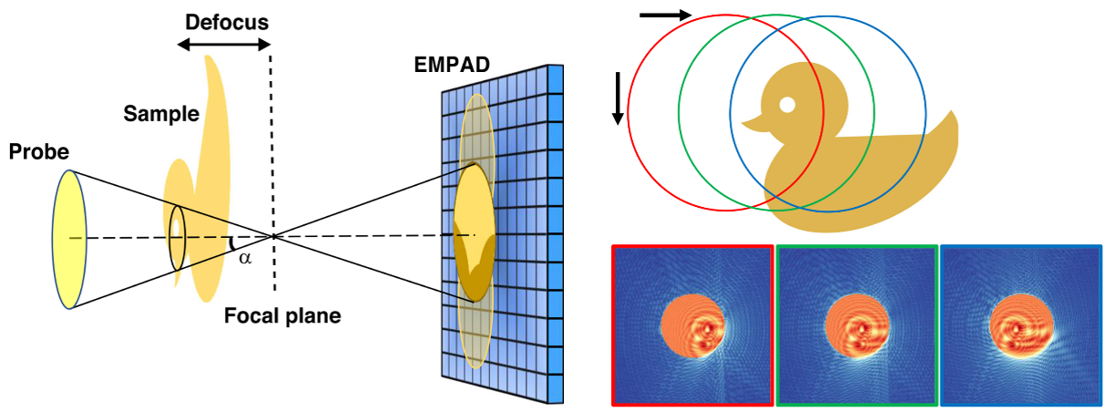
Figure 2: Experimental setup and simulation results, where the electron probe is focused downstream from the sample at the distance defined by the defocus value. The resulting diffraction pattern is collected on the detector (EMPAD) further downstream. See Highlight #27 for further details.
Data DOI: https://doi.org/10.34863/G4WA-0J57.
No. of Downloads: 427
Importance of Achievement: Demonstration of extremely low-dose imaging by electron ptychography (Figure 2), enabling the study of electron radiation-sensitive materials including biological macromolecules.
Associated Publication: Z. Chen, M. Odstrcil, Y. Jiang, Y. Han, M.-H. Chiu, L.-J. Li, and D.A. Muller“Mixed-state electron ptychography enables sub-angstrom resolution imaging with picometer precision at low dose,” Nature Communications 11, 2994 (2020). Publication DOI: 10.1038/s41467-020-16688-6.
Example C: Reaching the ultimate resolution limit
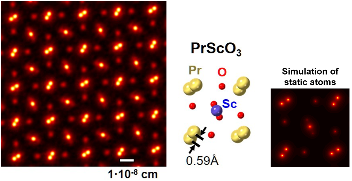
Figure 3: Microscopy image of PrScO3B, the corresponding crystal structure and an imaging simulation with static atoms, showing almost no blurring. See Highlight #42 for further details.
Data DOI: https://doi.org/10.34863/ssmm-2j11.
No. of Downloads: 208
Importance of Achievement: The Highest Resolution Microscope, enabled by a new detector technology, reaches an ultimate resolution limit – the vibrations of atoms themselves (Figure 3).
Associated Publication: Z. Chen, Y. Jiang, Y.-T. Shao, M.E. Holtz, M. Odstrčil, M. Guizar-Sicairos, I. Hanke, S. Ganschow, D.G. Schlom, and D.A. Muller, "Electron Ptychography Achieves Atomic-Resolution Limits Set by Lattice Vibrations," Science 372, 826-831 (2021). Publication DOI: 10.1126/science.abg2533.